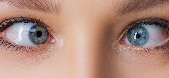
Keratoconus: how to recognize symptoms and causes

The editorial staff of Emianopsia.com has the pleasure of hosting Dr Chiara Spatafora: graduated in Orthoptics and Ophthalmological Assistance with additional qualification in optics. Dr Spatafora will give us an insight into keratoconus.
How can we define keratoconus?
This disease manifests as a dystrophy of the cornea, usually bilateral (about 96% of cases), but develops differently between the two eyes.
In most cases it is diagnosed during adolescence, rarely at the age of 25-30 years, and tends to increase up to 40 years, when it usually stabilizes.
Causes of this degenerative pathology of the cornea
It is a multifactorial pathology, where both genetic and environmental factors are involved:
- in some patients, autosomal dominant heredity with incomplete penetrance has been found, at least 10 mutated genes have been identified that could promote its occurrence.
- as for environmental factors, exposure from an early age to UV radiation and some behavioural factors such as “eye rubbing” (typical in children with allergies) determine an additional source of risk.
It can be present in more complex situations such as:
- Down’s syndrome;
- autoimmune diseases of connective tissue;;
- pathologies, such as Maarfan syndrome.
Signs and symptoms of keratoconus
This disease is characterized by a thinning of the stroma, the third layer that makes up the cornea: this alteration is often determined by a reduction in the synthesis of collagen by keratocytes and by the decrease and degeneration of the lamellae, creating a global deterioration in the quality of this connective tissue.
The disease can progress over time or be stationary (as in the case of the whipped keratoconus, that manifests itself mainly only in the form of irregular astigmatism).
Nonetheless, some clinical signs help us identify said pathology:
- corneal deformity: the cornea takes the characteristic form of a “cone” because of its thinning;
- astigmatism at the refractive level: it manifests following alterations on the corneal surface, generated by the variation of the corneal curvature radius. Astigmatism is often associated with myopia and, at the initial stage, this disease could be mistaken for a simple refractive defect. This pathology causes irregular astigmatism, that can be compensated with glasses only in the early stages, and is described by the patient as blurred and distorted vision;
- Munson’s sign: another characteristic sign that consists in the distortion of the lower eyelid, visible in the slit lamp, in the initial stages and are visible even without instruments in the more advanced stages;
- Vogt’s striae: these are stromal striae similar to scratches, often vertical;
- Fleischer rings: accumulation of hemosiderin pigment, an iron deposit located in the corneal epithelium; these rings can be yellow-brown and are visible using a slit lamp. Usually the more advanced the disease is, the thinner they become.
How is the diagnosis made?
Diagnosis and follow up require accurate eye examinations:
- measurement of visual acuity;;
- corneal topography: to map the anterior face of the cornea;
- corneal tomography: used to study the anterior and posterior side of the cornea;
- pachymetry: used to measure corneal thickness (under normal conditions the central thickness is 520-540 µm);
- corneal aberrometry;;
- inspection using a slit lamp.
Over the years, various classifications have been made:
- Amsler: this is the simplest classification, based only on the increase in the radius of curvature of the cornea;
- Krumeich: this classification embraces more parameters like myopia and astigmatism, the presence of scarred tissue ( aka Strie of Vogte ), and corneal thickness, which is measured with the pachymetry. According to the different stages of this illness, different medical interventions can be carried out.
Treatments and cures of keratoconus
In the initial stages, following a thorough eye examination with an ophthalmologist and a pachymeter, in case visual acuity is still detected, we proceed, initially, with optical aids such as glasses and soft contact lenses. These aids, of course, cannot stop the progression of the disease, but are helpful to correct blurred vision.
Photo selective lenses have also proven to be useful for protection against UV rays and helping younger subjects in not wrinkling their eyes.
The Corneal Cross-linking
In the initial forms of keratoconus, ( forms that will inevitably progress and which are not yet stabilizing ), it is possible to intervene with the Cross-linking corneal in order to preserve a 6-7/10 vision.
Cross-linking is a therapy that can slow down the evolution of the disease: usually the most indicated patients to undergo treatment are 15 to 25 years old, who do not show have Vogt’s striae yet, and with good visual acuity and, more importantly, a corneal tissue thickness greater than 400µm.
Cross-linking epi-off
This medical intervention is performed in the operating room, in local anaesthesia. First a corneal de-epithelization is performed (in the epi-off treatment) which then involves the removal of a thin layer of the epithelium; then riboflavin is administered, ( vitamin B2 complex that will be more easily absorbed as a result of the removal of the epithelial layer).
Riboflavin will then be irradiated by UVA: the radiation will increase the collagen present in the stroma, thus joining the cross-links (cross-linking) and blocking the progression. The surgery lasts about half an hour, after which the patient must wear therapeutic contact lens for at least 5 days.
Cross-linking epi-on
In addition to the “epi-off” treatment that has proven to be more and more promising in young subjects, cross-linking “epi-on” has also been experimented preserving the epithelium layer. Albeit being less invasive and painful, this procedure has proven less effective, since riboflavin fails to penetrate adequately at the level of the stroma. For this reason, this intervention is used only in certain cases, such as patients with a very thin cornea.
Hard contact lenses
In cases where this pathology has reached a progressive state, in which glasses and soft contact lenses can no longer correct the visual defect, the use of rigid contact lenses is advised. These lenses are designed to adapt to the specific corneal profile of the patient but, again, contact lenses do not have a therapeutic effect on the course of the disease.
Specific surgeries
In cases where even RGP contact lenses cannot improve visual acuity there are specific surgical interventions such as the introduction of intrastromal rings to strengthen the cornea and thus allowing the use of contact lenses.
Finally, in the most severe forms of keratoconus, cornea transplantation is used.
We thank Dr Spatafora for her article and, if you are interested in exploring this topic, check out this article: Eye diseases: the situation in Italy.
Bibliography
- Aldo Caporossi – Oftalmologia | Piccin-Nuova Libraia. 2017
Talk to a visual rehabilitation specialist now
Contact us without obligation to receive information.
Our Specialists will respond to you Monday through Friday from 9:00 a.m. to 5:00 p.m. (CET).
You are free to reproduce this article but you must cite: emianopsia.com, title and link.
You may not use the material for commercial purposes or modify the article to create derivative works.
Read the full Creative Commons license terms at this page.
You may also be interested in...

Color perception and diagnostic tests
Color perception greatly enriches in detail what we observe but can sometimes function abnormally. We learn about the psychophysical process of color vision, causes of perceptual deficits, and specific tests.


Stuttering: what is it, symptoms, and how to deal with it
Stuttering: what it is, how to distinguish symptoms, and how to improve quality of life.

Squint: types, symptoms and treatments
The causes of this problem are numerous, each of which contributes to describe the characteristic features of the various types of squint.


Attention deficit and hyperactivity: how to recognize it?
Attention deficit and hyperactivity disorder may be due to a multiplicity of factors, among which the most likely are genetic.

Diplopia: what are the causes and cures
Causes, symptoms and treatment of “Double Vision” or Diplopia.

Neurodevelopmental disorders: what do they entail?
Neurodevelopmental disorders make their onset in childhood and may persist into adulthood.