Diplopia: what are the causes and cures

The editorial staff of Emianopsia has the pleasure of hosting Dr Valeria Russolillo, Orthoptist Assistant of Ophthalmology. Dr Russolillo proposes a study on diplopia.
How can we define diplopia?
This issue entails a peculiar perception of reality: the patient seems to have two distinct images of a single object. We speak of monocular diplopia when this is also present by examining each eye individually. In these cases, the root of said issue is to be found in pathologies due to one of the structures of the eyeball such as:
- cataracts
- maculopathy
Conversely, this deficit is called binocular when it is caused by dysfunctions of the oculomotor system; in these cases the “double vision” disappears at the occlusion of one of the two eyes. The binocular diplopia is the one of greatest neuroophthalmological interest and is the most manifested form in the general population.
Here is a short video published by the Fondazione I.R.C.C.S. Istituto Neurologico Carlo Besta in which an overview of this issue is given.
Prof. Giuseppe Pinter Lauria (Fondazione I.R.C.C.S. – Istituto Neurologico Carlo Besta) explains diplopia and the synergy between neurologists and ophthalmologists.
Causes of diplopia
A common cause is the decompensation of a pre-existing squint in paediatric age, this is due to the progressive loss of the fusional reserves of compensation of heterophoria until then well controlled. In the rarest cases, however, it can occur because of … therapies not properly conducted.
This disorder is also the alarm bell of numerous neuroophthalmological diseases; it may occur as a result of paralysis or paresis of “central” eye motility, or ophthalmology manifested during inflammatory lesions and acute vascular such as cerebral stroke and TIA. Diplopia may also be the symptom of the onset of other demyelinating diseases of the central nervous system (such as multiple sclerosis) and autoimmune system (such as myasthenia).
When this problem arises from peripheral paralysis of the common oculomotor, trochlear and abducent nerves ( which regulate and contribute to maintaining the alignment of the visual axes ), it is attributable to the presence of systemic pathologies, such as diabetes and hypertension. Other triggers may be due to:
- tumour neoformations
- intracranial aneurysms
- endocranial hypertension.
Patients may also develop these deficiencies:
- post-surgical retinal detachment
- orbital injuries such as, for instance, trauma to the orbit, with muscle imprisonment
- following inflammatory thyroid dysfunction (particularly Basedow’s disease).
The symptoms
The “double vision” is the only symptom that is reported by the patient during an orthoptic evaluation; moreover the connected subjective symptomatology is very variable and sometimes inconsistent with what the orthoptist can objectively evaluate, through a large test battery.
This is due to the presence of several factors that contribute to change the representation in the space of the second image such as:
- the direction of gaze;
- the presence of a mixed vertical, horizontal or cyclotorsion squint
- refractive defects (not corrected)
- concurrent eye disease.
Not to be underestimated is the psychological perception of their state of visual health, for this reason we cannot disregard to listen carefully to what the patient reports, as to carry out an accurate medical history.
The ocular symptomatology that is associated with this disorder is related to the type of squint: if it is a divergent squint, patients initially report feeling blurry vision, “seeing through a water bubble” and then move on to see two very distinct images in presence of strong or neon lights, with a continuous feeling of glare.
In other cases we can find a patient with vertical diplopia present exclusively when looking up or down, symptom which increases when tilting the head; in these cases, also taking the stairs might constitute a problem, because of the double vision.
In the convergent squint (from paralysis or paresis of the sixth cranial nerve) the patient complains of diplopia only in the look from afar, therefore he is invalidated to the guide and in all the activities at a distance. In this case, a surgical evaluation is recommended in a short time, proposing the use of botulinum toxin to solve this clinical issue.
Therapy
As for the therapy, first it is important to transmit to the patient the basic information necessary to understand this visual discomfort that cannot be solved immediately, and with which you must undertake a long path of cohabitation and monitoring.
As already mentioned above, if the cause of this disorder is attributable to a glycaemic or hypertensive peak, the orthoptist needs to refer the patient to their general practitioner and specialist in order to resolve or stabilize the patient’s personal situation and re-evaluate it in the light of a new therapy and any diagnostic investigations (such as, for example, magnetic resonance imaging with regard to the need to diagnose certain neurological pathologies).
The patient’s squint is then re-evaluated by measuring it objectively in prismatic dioptres (dp) to verify how this has changed over time. In the literature, in most cases, there is a considerable initial improvement, followed by a modest progress and a stationary period in the next 10-12 months, before which surgery is not advised
The oculomotor picture or a slight diplopia (with a small angle of squint less than 10-15 dp) can be resolved with prismatic lenses, which can be applied on their own or incorporated into the lenses of the glasses. Depending on the discomfort reported by the patient, prismatic lenses can also be used temporarily while waiting for the angle of squint to be reduced.
Another option (only under the advice of qualified staff) can be using filters customized for the patient, to exclude the second image produced by the eye affected by paralysis or paresis. In any case, it is not recommended to close the eye with a bandage or similar materials and deprive it of receiving light. The orthoptist is a rehabilitation professional and as such their task is to bring the eye to look forward.
Bibliography
- Guida alla professione di ortottista Litografia A. Trischitta – Messina Maggio 2009
- Visione binoculare e motilità oculare, teoria e trattamento dello strabismo, gunter k. Von Noorden
- Diplopia S. Bidot, D. Biotti, C. Vignal-Clermont
Talk to a visual rehabilitation specialist now
Contact us without obligation to receive information.
Our Specialists will respond to you Monday through Friday from 9:00 a.m. to 5:00 p.m. (CET).
You are free to reproduce this article but you must cite: emianopsia.com, title and link.
You may not use the material for commercial purposes or modify the article to create derivative works.
Read the full Creative Commons license terms at this page.
You may also be interested in...
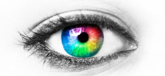
Color perception and diagnostic tests
Color perception greatly enriches in detail what we observe but can sometimes function abnormally. We learn about the psychophysical process of color vision, causes of perceptual deficits, and specific tests.


Stuttering: what is it, symptoms, and how to deal with it
Stuttering: what it is, how to distinguish symptoms, and how to improve quality of life.

Squint: types, symptoms and treatments
The causes of this problem are numerous, each of which contributes to describe the characteristic features of the various types of squint.

Attention deficit and hyperactivity: how to recognize it?
Attention deficit and hyperactivity disorder may be due to a multiplicity of factors, among which the most likely are genetic.


Neurodevelopmental disorders: what do they entail?
Neurodevelopmental disorders make their onset in childhood and may persist into adulthood.

Verbal language in autism: how to communicate?
What are the communication problems involving verbal language in autism? Here’s how to act on this issue.