Visual rehabilitation: promises and hopes

We are here today to present a pilot study (the VISION study) aimed at comparing three kinds of treatment for the recovery of patients in which, following a stroke, homonymous hemianopsia has started to manifest. The three treatment methodologies compared are visual rehabilitation, external optical aids and the standard treatment procedure (focused mainly on advice).
Before we get started here is a video from Santo Stefano Riabilitazione.
What caused this loss of sight?
What caused this loss of sight?
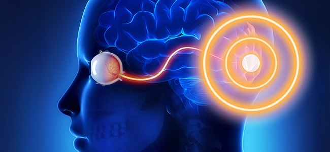
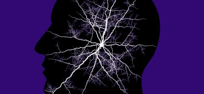
Stroke is one of the triggering circumstances (to discover all the other causes check our article “Hemianopsia: symptoms and causes”) of this visual deficit that involves the loss of half of the visual field. The symptomatology that damages the right or left visual field takes the name of homonymous hemianopsia (to discover all the other types of hemianopsia and learn how to distinguish them, check out this article “Hemianopsia homonymous: how to distinguish it?”).
What are the issues correlated with reduced vision?
It is immediately understandable that such a visual deficit entails a series of problems and limitations in the everyday life of hemianoptic subjects. From difficulty in reading and movement to the impossibility of driving, these are only some of the problems that hemianopsia brings on, resulting in a series of collateral problems such as depression and a sense of inadequacy (to further learn about these topics read “Hemianopsia issues: what are they?”).
Visual rehabilitation: there is still a long way to go
This type of methodology is gradually gaining popularity in the treatment process aimed at the recovery of people who have hemianopsia following a stroke. Its limited application within the departments of the national health system, unfortunately, gives too few indications about rehabilitation effectiveness and economic sustainability. Therefore, it is necessary to test this kind of rehabilitation through studies and clinical research.
VISION Studio: methods and applications
Through the random selection of testers, the VISION study compared:
- Visual rehabilitation through visual research exercises: testers were trained to perform more accurate saccade (eye micromovements) to effectively inspect the side involved in visual impairment.: i tester venivano allenati a compiere saccadi (micromovimenti oculari) più accurate volte a ispezionare efficacemente il lato coinvolto nel deficit visivo.
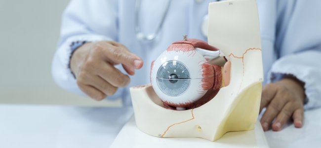
- External optical aids: treatment that makes use of prismatic glasses, in this case, “monocular” (having prismatic lenses on a single eye). With the help of these lenses, images from the blind field are projected towards the non-damaged visual area.
- Standard treatment..
The VISION study, after a screening phase of the testers that took place between 2011 and 2013 and that involved 87 candidates, then divided the suitable candidates into three groups (each focused on a type of treatment) random way:
- Group A: focused on prismatic glasses.
- Group B: focused on visual rehabilitation.
- Group C: focused on standard support.
A first evaluation of the study we have the following report:2:
- 69% of the testers belonging to group A, that is having recourse to prismatic lenses, had contraindications (usually headaches).
- Only 7% of the candidates in Group B had contraindications (fatigue and migraine).
- No contraindications were recorded for group C members.
Improvements in the field of vision were:3:
- 5% for group A.
- 8% for group B.
- 3.5% for group C.
VISION study: the conclusions
Through this study, we have been able to show the improvements, albeit still small, and for medical research to now focus on visual training, bypassing the complications that prismatic lenses carry and that could prove to be a real disincentive for the patient to want to proceed with this type of treatment.
Bibliography
- Stroke Association | Can Rehabilitation help with visual field visual loss after stroke? Giugno 2016
- Ibid.
- Ibid.
Publications
- Rowe, Fiona J., Marion Walker, Janet Rockliffe, Alex Pollock, Carmel Noonan, Claire Howard, Richard Glendinning, Rachel Feechan, and Jim Currie. “Care provision for poststroke visual impairment.” Journal of Stroke and Cerebrovascular Diseases 24, no. 6 (2015): 1131-1144.
- Rowe FJ, Barton PG, Bedson E, et al. A randomised controlled trial to compare the clinical and cost-effectiveness of prism glasses, visual
- search training and standard care in patients with hemianopia following stroke: a protocol. BMJ Open. 2014;4(7):e005885. doi:10.1136/
- bmjopen-2014-005885.
- Rowe FJ, Conroy EJ, Barton PG, et al. A randomised controlled trial of treatment for post-stroke homonymous hemianopia: screening and recruitment. Neuro-ophthalmology. 2016;40(1), 1-10. doi: 10.3109/0165 8107.2015.1126288.
Talk to a visual rehabilitation specialist now
Contact us without obligation to receive information.
Our Specialists will respond to you Monday through Friday from 9:00 a.m. to 5:00 p.m. (CET).
You are free to reproduce this article but you must cite: emianopsia.com, title and link.
You may not use the material for commercial purposes or modify the article to create derivative works.
Read the full Creative Commons license terms at this page.
You may also be interested in...
Stroke: what happens to eyesight?
This article investigates visual impairment caused by stroke, treatments for rehabilitation of hemianopia and visual neglect, risk factors, and prevention.
Hemianopia in relation to other pathologies
Hemianopsia is often the first onset of severe brain disease (e.g., aneurysms, tumors, and strokes). Let us review some rare cases of hemianopsia related to less common diseases.


Treatments for patients with hemianopia
Going downstairs, grasping objects, reading and writing are some of the difficulties that patients with hemianopsia face on a daily basis. Let’s find out how rehabilitation plays an important role in visual recovery.


Cerebral ischemia: what it is, how to recognize symptoms
Cerebral ischemia indicates a distressed status of an area of the brain. Here’s who it affects and the symptoms not to underestimate.
Stroke: symptoms, consequences, and rehabilitation
Cerebral stroke is the third cause of death in Italy. How can we recognize the symptoms and how does rehabilitation take place?


Neglect: what is it, how to recognize it and how to cure it
Neglect is a visual deficit characterized by complete disinterest towards one side of the visual field.
