Hemianopia in relation to other pathologies

Hemianopias are unilateral or bilateral Visual Field defects resulting from chiasmatic or retrochiasmatic lesions.
They are distinguished into:
- Heteronymous (binasal or bitemporal) o Homonymous (lateral or altitudinal)
- Complete or Incomplete (quadrant – quadrantanopia)
- Congruous or Incongruous
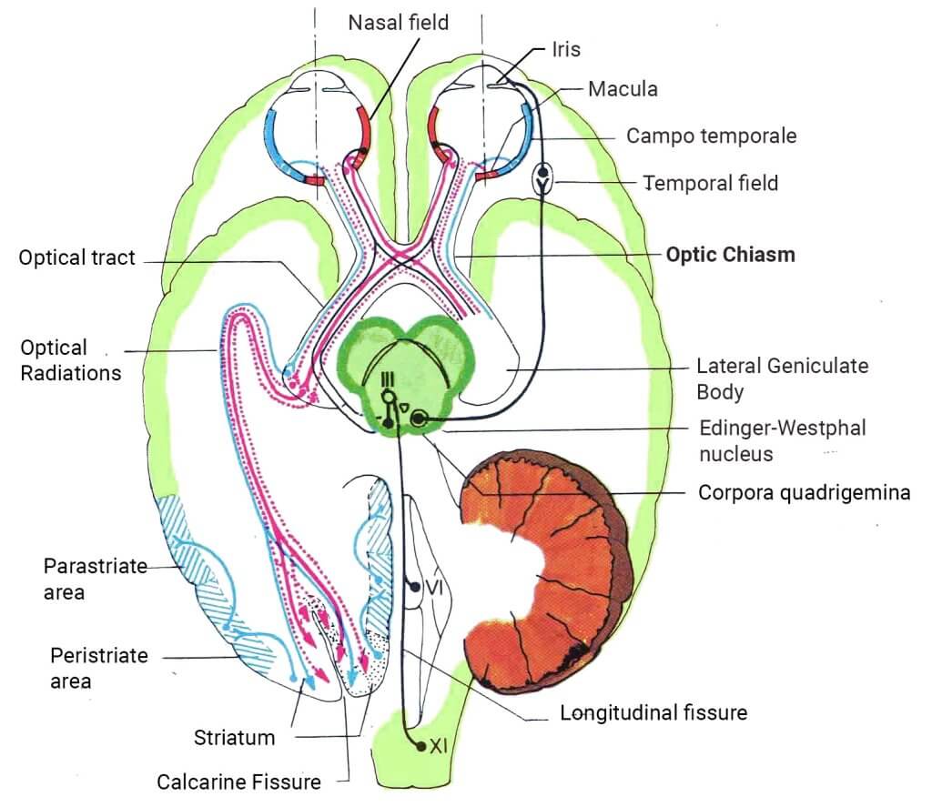
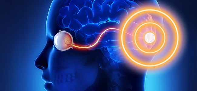
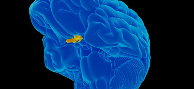
The optic chiasm (see Figure 1), or the junction through which direct visual information passes to the brain, can be affected by clinical pictures that result in its alteration. The involvement of the optic chiasm in many pathologies is mainly due to the small space present at the level of the suprasellar region.
Chiasmatic pathology can be related to the following pathological situations:
- tumors in 25% of cases, such as:
- pituitary adenomas (50%)
- cranipharyngiomas (25%)
- meningiomas (10%)
- gliomas and metastases of systemic tumors (7%)
- aneurysms
- demyelination
- vasculitis.
Tumors often result in chiasmatic suffering due to compressive or infiltrative action; they mainly result in visual field alteration:
- pituitary adenomas result in bitemporal hemianopsia beginning superiorly
- craniopharyngiomas are characterized by bitemporal hemianopsia beginning inferiorly
- meningiomas originating at the level of the tubercle of the sella determine junctional scotoma.
Optic tract pathology affects those portions of the optic pathway, post or retro-chiasmatic and is characterized by incongruous homonymous hemianopsia, counter lateral to the side of the lesion.
The optic tract ends its course by reaching the visual cortex, located at the level of the occipital lobe, along the margins of the calcarine scissure.
During its path a series of radiations arise, that is, a series of beams directed at certain areas of the cortex, and depending on the radiation affected, characteristic visual field alterations will occur.
- if the one affected is the main radiation, there will be complete bitemporal hemianopsia
- usually a bitemporal hemianopsia is to be referred to a complete chiasmatic lesion
- a right nasal hemianopsia is to be referred to a perichiasmatic lesion;
- a right homonymous hemianopsia is to be referred to a complete left optic tract lesion or left optic radiation
- a right homonymous hemianopsia with macular sparing to a calcarine lesion.
The following will be some rare cases of manifestation of hemianopsia related to less common diseases than those described above, on which landmark clinical studies have been performed.
Monocular nasal hemianopsia caused by traumatic optic neuropathy
The case is that of a 41-year-old male who was punched in the head during a fight and then suffered painless blurred vision in his left eye after a mild traumatic brain injury. 1
An ophthalmic examination revealed conjunctival chemosis, periorbital hematoma, and a related afferent pupillary defect in the left eye. Automated perimetry indicated the presence of left-sided nasal hemianopsia along the vertical meridian.
In this case, the monocular nasal hemianopsia was probably due to ischemic changes resulting from impairment of the prechiasmal arterial anastomotic network or indirect injury to the lateral prechiasmal nerve fiber. An ophthalmologic examination after treatment (systemic therapy with 1 gram methylprednisolone intravenously daily for 3 days) indicated that there was no obvious improvement in relative afferent pupillary defect, better corrected visual acuity and color sense.
A second set of automated perimetry results showed no change after 3 months.
Copyright © 2020 the Author(s). Published by Wolters Kluwer Health, Inc. 1
Permanent and complete bilateral homonymous hemianopsia with preservation of central vision
Rare example of a patient who developed permanent and complete bilateral homonymous hemianopsia with preservation of central vision after transtentorial herniation secondary to subdural hematoma. 2
The patient developed severe amnsesia (memory loss) and retrograde amnesia and spatial disorientation; he acted as if he were blind but could recognize a small pin on the floor.
The spatial disorientation persisted, and although he spent many days in the hospital, the patient never learned to go from his room to the bathroom. Also, while in his room, he could not find his own bed. Another notable deficit was complete memory loss of recent events. These severe neurological deficits remained essentially unchanged during a four-year follow-up period. The bilateral homonymous hemianopsia in the case reported here is of particular interest because of its completeness and because of the full preservation of central vision with normal visual acuity in the central 10-20 degrees of both eyes.
Traumatic bitemporal hemianopsia with macular sparing
Traumatic chiasma syndrome is a rare complication of closed head injury. It often presents as bitemporal hemianopsia and may be associated with other neurological signs. 3
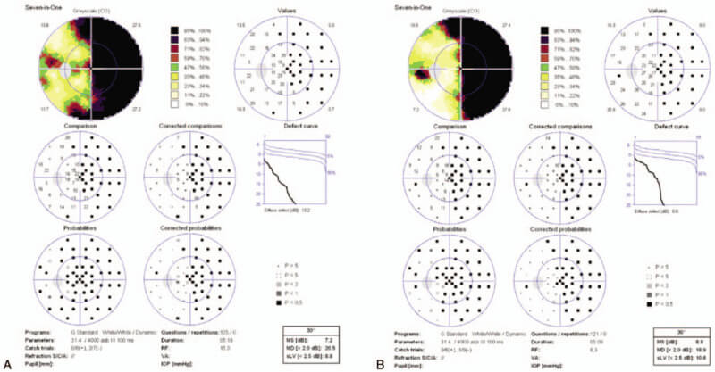
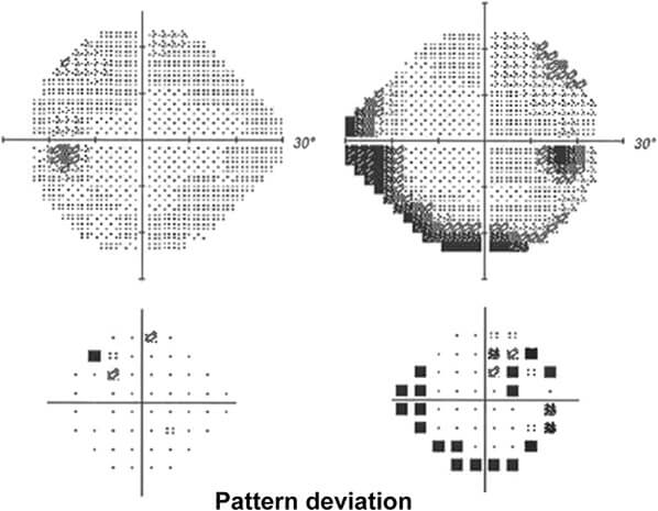
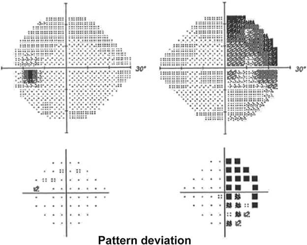
Some authors report the case of a 47-year-old man who had suffered severe frontal head trauma following an automobile accident that resulted in multiple skull fractures and prolonged loss of consciousness. He was subsequently diagnosed with traumatic chiasm syndrome. Tangent field testing revealed bitemporal hemianopsias with some macular sparing. Macular sparing was not found in the central 30-2 pattern of Humphrey’s visual field test.
Case of monocular temporal hemianopsia
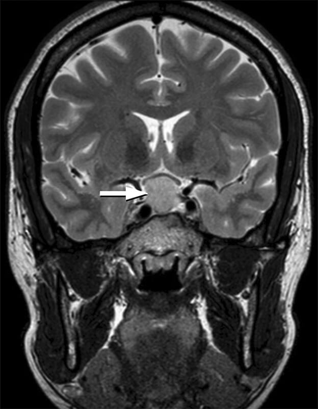
A 35-year-old first-pregnancy woman presented to the eye care emergency room with reduced visual acuity in her right eye. Humphrey’s visual field test showed monocular temporal hemianopsia in the right eye before delivery. An MRI after delivery revealed a widely symmetric pituitary macroadenoma with chiasmal compression.
This is a rare case of pituitary macroadenoma (benign tumor affecting the pituitary gland larger than one centimeter in size), especially when the tumor is largely symmetrical. 4
Journal of Neuro-Ophthalmology41(2):e267-e268, June 2021. 4
Journal of Neuro-Ophthalmology41(2):e267-e268, June 2021. 4
Journal of Neuro-Ophthalmology41(2):e267-e268, June 2021. 4
Homonymous hemianopsia as a consequence of diabetic ketoacidosis
Neurological complications of hyperglycemia are common. Cases of seizures and hemianopsias related to non-ketone hyperglycemia have already been reported, but they are rare in diabetic keto acidosis. 4
Neurological complications of hyperglycemia are diverse, including choreoathetosis (neurological syndrome characterized by the onset of involuntary movements), hemiballismus (movement disorder affecting a limb), dysphagia (difficulty swallowing), seizures, and coma. Various types of seizures (occipital, focal or complex partial) have been reported together with hemianopsias in the context of NKHH (nonketotic hyperosmolar hyperglycemia or hyperglycemic hyperosmolar state).
Emianopsia: effetto di un’altra patologia
Treatment of hemianopsia basically aims to remove the causes of the damage, for example, by removing a tumor mass. Unfortunately, in some cases, even if this is surgically possible, the damage is irreversible and it is therefore impossible to “recover” the lost portion of the visual field.
Thus, hemianopsia must be considered the effect of another pathology: it is often the first onset of a serious brain disease (aneurysms, tumors, stroke, etc.) that may not show signs until after the loss of the visual field. 7
Rehabilitation, however, plays an important role in patients with hemianopsias. Scientific studies have shown that an approach based on audiovisual visual field stimulation induces lasting improvement in visual field deficits. 8
Article references
- Lin HL, Yen JC. Acute monocular nasal hemianopia following a mild traumatic brain injury: A case report. Medicine (Baltimore). 2020 Jul 24;99(30):e21352. doi: 10.1097/MD.0000000000021352. PMID: 32791737; PMCID: PMC7386976.
- Soza M, Tagle P, Kirkham T, Court J. Bilateral homonymous hemianopia with sparing of central vision after subdural hematoma. Can J Neurol Sci. 1987 May;14(2):153-5. doi: 10.1017/s0317167100026299. PMID: 3607617.
- Soza M, Tagle P, Kirkham T, Court J. Bilateral homonymous hemianopia with sparing of central vision after subdural hematoma. Can J Neurol Sci. 1987 May;14(2):153-5. doi: 10.1017/s0317167100026299. PMID: 3607617.
- Pituitary Macroadenoma Presenting With Monocular Temporal Hemianopia – Gupta A, Deshmukh M, Palexas G. – J Neuroophthalmol. 2021 Jun 1;41(2):e267-e268.
doi: 10.1097/WNO.0000000000001119. PMID: 33136669. - J Med Assoc Thai. 2005 Nov;88 Suppl 9:S126-30.
- Chukwudelunzu FE, Fugoso L. Diabetic Ketoacidosis Causing Transient Homonymous Hemianopia and Generalized Seizure: A Case Report and Literature Review. WMJ. 2023 May;122(2):127-130. PMID: 37141479.
- Malattie oculari: Emianopsia – IAPB Italia Onlus: Agenzia Internazionale per la Prevenzione della Cecità
- Nadia Bolognini, Fabrizio Rasi, Michela Coccia, Elisabetta Làdavas, Visual search improvement in hemianopic patients after audio-visual stimulation, Brain, Volume 128, Issue 12, December 2005, Pages 2830–2842, https://doi.org/10.1093/brain/awh656
Bibliography
- Homonymous Hemianopsia – Adam Wolberg; Nidhi Kapoor.
- Hemianopsia – Jonah Ruddy; Alfonso C. Cardenas.
- Neuroftalmologia – Francesco Carta, Arturo Carta – edizioni Monduzzi , 2009
Talk to a visual rehabilitation specialist now
Contact us without obligation to receive information.
Our Specialists will respond to you Monday through Friday from 9:00 a.m. to 5:00 p.m. (CET).
You are free to reproduce this article but you must cite: emianopsia.com, title and link.
You may not use the material for commercial purposes or modify the article to create derivative works.
Read the full Creative Commons license terms at this page.
You may also be interested in...

The other half of the world: interview with someone living with hemianopia
In this interview, Marco shares his experience with courage and honesty, opening up without holding back. It’s an invitation not to give in to what’s missing, but to rediscover the value of every piece that remains.

Stroke: what happens to eyesight?
This article investigates visual impairment caused by stroke, treatments for rehabilitation of hemianopia and visual neglect, risk factors, and prevention.

Treatments for patients with hemianopia
Going downstairs, grasping objects, reading and writing are some of the difficulties that patients with hemianopsia face on a daily basis. Let’s find out how rehabilitation plays an important role in visual recovery.


Types of Hemianopias
Let’s clarify the types of hemianopias by simulating the vision of a person with this important visual field deficit.

Hemianopsia and diseases of the optic chiasma
Lesions of the optic chiasm can cause various visual field alterations: the most common alteration is bitemporal heteronymous hemianopsia.

Hemianopsia: neuroscience and neuropsychological rehabilitation
The rehabilitation of hemianopsia requires multidisciplinary teamwork and the creation of an customized rehabilitation project.
