Hemianopsia: causes and characteristics of this visual impairment

Our editorial staff is pleased to host in this virtual space Dr. Catalano: an ophthalmic surgeon, a graduate of the University of Catanzaro’s “Magna Graecia” School of Medicine and an expert in the diagnosis and treatment of retinal, corneal and glaucoma diseases. Dr. Catalano provides us with an in-depth look at hemianopsia: causes and characteristics, providing readers with expertise and knowledge that can shed light on this peculiar visual impairment.
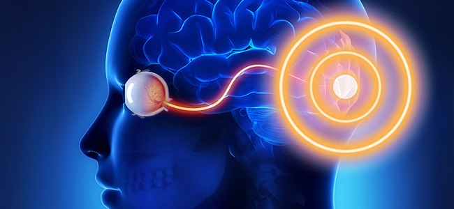
Operation and components of optical pathways
This visual impairment consists of partial damage to half of the visual field and can be unilateral or bilateral, that is, involving one eye or both. Any damage from the cerebral cortex to the retina can cause visual field damage.
Optical pathways consist of three functional components:
- Optics: from cornea to retina.
- Retinocortical:from retina to primary visual cortex.
- Integrative: from primary visual cortex to vision-related parietal and temporal cortex.
The optical component carries the light signal to the retina. In the retinocortical section, the signal reaches the photoreceptors , which transform the light signal into an electrical signal and so it is transmitted to the visual cortex for elemental encoding.
In the integrative portion, the occipitoparietal pathway integrates motion and depth information into spatial perception. The occipitotemporal pathway integrates shape and color information into recognizable symbols.
Homonymous hemianopsia: from what does it originate?
Generally, the hemianopsia has a chiasmatic or postchiasmatic origin, that is, a lesion present in the section where nerve fibers from the optic nerve cross or have already divided and passed into the opposite hemisphere. In fact, nerve fibers from both visual hemifields are present in these areas because the outermost portion, from the temporal hemiretina, does not decussate (45 percent), while nerve fibers from the nasal hemiretina decussate (55 percent).
Hemanopsia: causes
Therefore, generally, the causes that can lead to this visual impairment are:
- Partial temporal optic nerve injury (unilateral nasal).
- Retinal ischemic phenomena that may give superior or inferior (unilateral) hemianopsia.
- Partial medial optic nerve injury (unilateral temporal).
- Retinal trauma (unilateral).
- Injury to the optic chiasm (bitemporal).
- Lesion to the optic tract (right or left homonymous).
What are the origins of the lesions from which this symptomatology arises?
These particular types of injuries that can go on to generate visual field loss can have the following origin:
- Ischemic.
- Traumatic.
- Compressive.
- Toxic.
In all of the above cases, neuronal damage occurs, leading to blindness of the affected area.
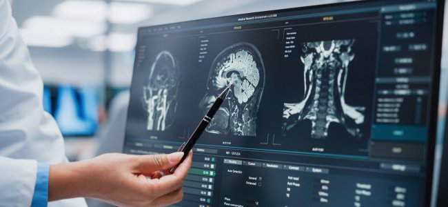
What are the tests we can use?
A very useful examination in cases of optic pathway injury is the computerized visual field. It is a subjective examination in that the patient must recognize a light stimulus and report it to the machine, which will subsequently translate the responses into an explanatory graphic reconstruction in addition to all the parameters useful to the ophthalmologist and neurologist for follow-up of the pathology.
Instead, objective examinations are the electrofunctional ones, in which a light stimulus is exhibited to the retina and through electrodes the amplitude and latency of the response is studied.
New hope comes from neurofunctional rehabilitation
Such injuries can lead to significant disability, limiting lifestyle, work and social activity with an impact on both the emotional state and, consequently, the patient’s overall health status. To learn more read our article Homonymous lateral hemianopsia: here are the issues.
While visual rehabilitation of such injuries was once considered unnecessary and superfluous, the evolution of rehabilitation techniques today can give many patients hope. Neurofunctional rehabilitation aims to strengthen healthy areas so that they can make up for the inabilities of damaged areas.
We thank Dr. Catalano for providing this insight intohemianopsia: causes and characteristics of what is a very peculiar symptomatology.
Acknowledgements
- Cover photo by Aditya Wardhana on Unsplash
Talk to a visual rehabilitation specialist now
Contact us without obligation to receive information.
Our Specialists will respond to you Monday through Friday from 9:00 a.m. to 5:00 p.m. (CET).
You are free to reproduce this article but you must cite: emianopsia.com, title and link.
You may not use the material for commercial purposes or modify the article to create derivative works.
Read the full Creative Commons license terms at this page.
You may also be interested in...

The other half of the world: interview with someone living with hemianopia
In this interview, Marco shares his experience with courage and honesty, opening up without holding back. It’s an invitation not to give in to what’s missing, but to rediscover the value of every piece that remains.

Stroke: what happens to eyesight?
This article investigates visual impairment caused by stroke, treatments for rehabilitation of hemianopia and visual neglect, risk factors, and prevention.
Color perception and diagnostic tests
Color perception greatly enriches in detail what we observe but can sometimes function abnormally. We learn about the psychophysical process of color vision, causes of perceptual deficits, and specific tests.

Hemianopia in relation to other pathologies
Hemianopsia is often the first onset of severe brain disease (e.g., aneurysms, tumors, and strokes). Let us review some rare cases of hemianopsia related to less common diseases.

Treatments for patients with hemianopia
Going downstairs, grasping objects, reading and writing are some of the difficulties that patients with hemianopsia face on a daily basis. Let’s find out how rehabilitation plays an important role in visual recovery.


Types of Hemianopias
Let’s clarify the types of hemianopias by simulating the vision of a person with this important visual field deficit.